

Coronary Angiography & Angioplasty
Coronary Angiography
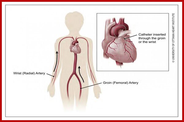
An angiography or angiogram is a special type of X-ray that allows your coronary arteries to be viewed and recorded on film. This X-ray allows your doctor to see if the blood vessels to your heart are clogged.CAG is usually performed from the hand (Radial Angioplasty) but sometime its done from groin too (Femoral Angioplasty).
Before Your Angiography
• Tell your doctor what medicines you take and any allergies you may have.
• Don’t eat or drink anything after midnight on the night before the procedure.
• Arrange for an adult family member or friend to drive you home.
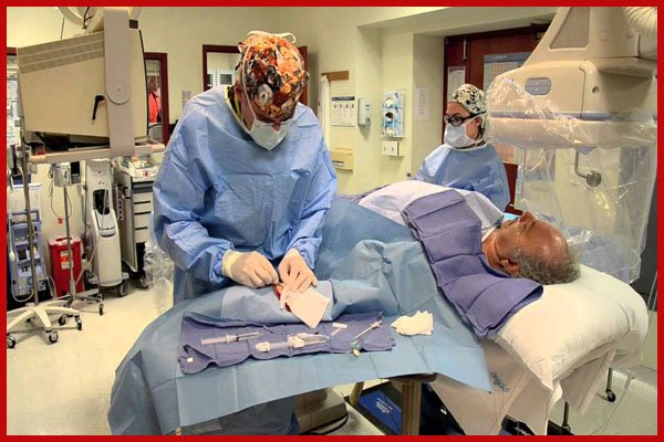
During Your Angiography
• A long, thin tube called a catheter is placed inside an artery in your groin or arm and guided into your heart.
• A contrast dye is injected through the catheter into your blood vessels or heart chambers.
• X-rays are taken to show clear photos of the inside of your heart and coronary arteries.
After Your Angiography
• You need to remain remain under observation for few hours.
• If the insertion site was in your groin, you may be required to lie down with your leg still for several hours.
• A nurse will check your blood pressure and the insertion site.
• You may be asked to drink fluid to help flush the contrast liquid out of your system.
• An adult family member or friend will need to drive you home from the hospital.
• It’s normal to find a small bruise or lump at the insertion site. These common side effects should disappear within a few weeks.
Carotid Angiography & Angioplasty
A carotid angioplasty is a minimally invasive technique used to widen a narrowed or obstructed artery. During the procedure, a long, thin tube called a catheter is inserted into the artery. This is used to move instruments through the artery to put the stent in place.
Before Inserting the Catheter
• The skin in the area of the insertion site is numbed with local anesthetic. A puncture is made in the femoral artery, a major artery in the groin.
• An introducing sheath (tube) is inserted into the puncture.
• The catheter is inserted into the sheath. Using X-rays as a guide, the doctor moves the catheter up the aorta, behind the heart and to the carotid artery.
During a Carotid Angioplasty
A filter or other protective device prevents pieces of plaque from being carried into the brain and causing a stroke. The catheter is used to place the unopened filter in the artery and advance it past the narrowed area. The filter is then opened. It remains in place for the whole procedure.
If narrowing is very severe, the artery may need to be widened or opened before the filter is put in place. This is done using balloon angioplasty.
• A tiny, uninflated balloon is first moved to the area that needs to be widened.
• The balloon is then inflated, pushing the artery open.
• After the procedure is done, the balloon is deflated and removed.
Placing the Stent
• The stent is moved to the site of the plaque.
• The catheter is then withdrawn, leaving the stent in place.
• The stent expands until it touches the plaque. Balloon angioplasty is then used to expand the stent fully and widen the artery.
• The balloon is withdrawn, leaving the stent in place to hold the artery open.
After an Angioplasty
An angiogram is taken and compared to the one that was taken at the beginning of the procedure. Once your doctor is satisfied with the result, the filter and other instruments are withdrawn. The groin insertion site is then closed. Your doctor will also discuss care and follow-up appointments with you.